There are at least 3,000 species of mites (Arachnida: Acari) living in close association with birds (Aves). The influence of mites on host survival and reproduction ranges from severely detrimental to mildly beneficial. Four orders of mites include species that are blood or tissue feeders on birds. While surveying mites from birds in Alberta, we became aware of the need for an inclusive illustrated key to identify the families and genera of blood and tissue feeding mites. We collected mites by washing birds and examining the filtrate. In total, 328 individual birds representing 123 species were examined. We used a camera-mounted compound microscope, Adobe Photoshop, and a drawing tablet to acquire and edit images. The resulting key covers 13 families and 14 genera of parasitic mites. It was created with non-acarologists in mind and can be easily used by anyone interested in identifying bird-associated mites.
Ptilonyssus bombycillae (Rhinonyssidae)
ex. Bohemian Waxwing (Bombycilla garrulus).
We collected approximately 600 Albertan birds contributed by the Alberta Fish & Wildlife Forensic Laboratory, the Royal Alberta Museum, waterfowl hunters, and colleagues at the University of Alberta. Our key is based on examination of washings from about 328 individual birds representing 123 species from 16 orders. There are 402 species of birds in Alberta, thus our sample represents 31% of Alberta’s avian fauna (Royal Alberta Museum 2005).
Birds were maintained at -20ºC until processing. Frozen birds were first thawed and then washed using the following method. The bird was placed in a suitably sized container, ranging from 4-18L, with a drop of dish detergent, enough 95% ethanol to soak the plumage of the bird, and enough water to submerge it. The sealed container was then shaken vigorously for five minutes. Particularly large birds were washed in a basin and thoroughly massaged while in the solution. Each bird was then removed from the container and rinsed thoroughly over a Fisher Scientific 53 µm mesh filter; large birds were rinsed over the washing basin. The washing liquid was filtered through the same 53 µm filter. The container and lid were rinsed thoroughly over the same filter as well. The filtrate was stored in 30 ml scintillation and snap cap vials. For approximately half of the birds that were washed, no particular attention was paid to flushing the respiratory passages to remove nasal mites. For the remainder we ensured that the nasal passages were flushed with 95% ethanol.
Washings were examined using Leica MZ16 and MZ6 dissection microscopes at 20-25x magnification. Blood and tissue feeding mites were removed, and most were cleared in 85% lactic acid for one to twenty four hours depending on the degree of original transparency. Mites were mounted in a polyvinyl alcohol medium (PVA) from Bioquip Products Inc. (6371A). Slides were cured on a slide warmer at about 40ºC for three to four days.
We made our identifications using a combination of synoptic literature (Krantz 1978; McDaniel 1979) and primary literature (Fain and Elsen 1967; Moss 1978; Pence 1975; Smiley 1970). Images of selected slide-mounted mites were taken with a Canon Powershot S40 digital camera mounted on a Leica DMLB compound microscope with DIC, at 200-400x magnification. Images were captured in the Canon Utilities Remote Capture program, version 2.2.0.11. We edited images using Adobe Photoshop CS version 8.0, Adobe Systems Inc. For a generalized representative of each of the four orders collected, selected mites were drawn using an Intuos 2 Graphics Tablet from WACOM Co., Ltd. and Adobe Photoshop CS version 8.0. Two representative mites were drawn from images taken as described above and the other two mites were redrawn from published literature, one from Smiley (1970) and the second from Moss (1968).
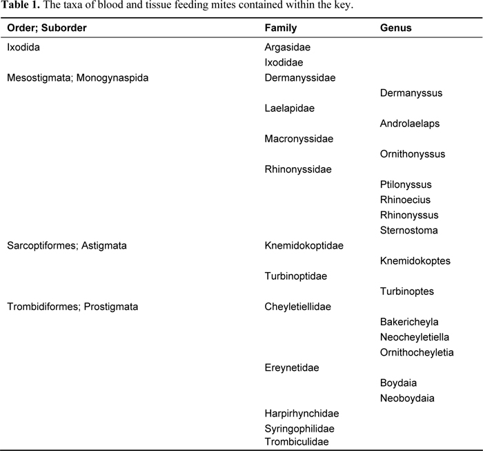
The key covers 14 genera from 13 families of blood and tissue feeding mites (Table 1). All taxa, with one exception (soft ticks, Argasidae), were collected from our specimens of Albertan bird specimens (Table 2). We collected blood and tissue feeding mites, from 38% of 328 individual bird specimens and 58% of 91 genera examined. Mites of the family Rhinonyssidae were collected from the most bird genera (25), followed by Harpirhynchidae (23 host genera) (Table 2). Harpirhynchids were not identified to genus due to time constraints. Host association records presented here are not intended to be exhaustive since examination of more bird specimens and taxa will undoubtedly reveal additional records.
Our key to parasitic mites associated with Albertan birds begins with brief section on feather mites, whose families are not covered in the key since feather mites are largely benign (Proctor and Owens 2000). Gaud and Atyeo (1996) developed synoptic keys to feather mite genera of the world. The body of our key has two main parts, the first to families and the second to genera, and is based primarily on adult females. Within the key there are links to labeled diagrams illustrating important morphological characteristics of the four orders of mites collected.

We particularly thank Bob McClymont and the Alberta Fish and Wildlife Forensic Laboratory, as well as the Royal Alberta Museum for providing us with numerous specimens. Dr. David Walter provided information and helpful criticism and Dr. Bruce Heming advice and editing. S. Grundke and S. Wojtkiw washed birds during the summer of 2003, and N. Romaniuk and M. Pedruski did the same during the summer of 2004. G. Williams and H. Shannon allowed us to use a few of their images in the key. We thank Dr. Al Shostak for his assistance in identifying ticks, as well as the use of his facilities. Lastly, we thank Wayne Roberts for providing us with some unique specimens.
We conducted this research with a scientific salvage permit to take migratory birds from Environment Canada (permit no. CWS03-A009), and a permit to salvage found dead wildlife (excluding endangered species) from Alberta Environment (permit no. 428 CN). This project was funded by an NSERC Discovery grant to HCP and an NSERC USRA to W. Knee.
Table 2. The taxa of blood and tissue feeding mites associated with Albertan birds, with the families and genera of hosts from which they were collected.
Benign Mites Commonly Associated with Birds:
There are approximately 2000 described species of feather mites (Astigmata) worldwide; most birds have feather mites. Feather mites belong to the superfamilies Analgoidea, Freyanoidea and Pterolichoidea. These mites are benign; hence they are excluded from this key. Not all astigmatans are feather mites; some are blood and tissue feeders. Here are a few species of feather mites collected from a Sora Rail, Porzana carolina Linnaeus.
To discriminate feather mites from other mites, feather mites always lack tarsal claws on at least legs 1 and 2, instead they have an elaborate ambulacrum on the end of the tarsi. For comparison, here is the ambulacrum and tarsal claws of legs 1 and 2 of a tissue feeding astigmatan nasal mite.
Key to the Families of Blood and Tissue Feeding Mites Known from Birds in Alberta (primarily based on females)
The identification of some mites, especially members of the order Mesostigmata, requires an identifier to discriminate between males and females. All mesostigmatan males have a prominent gonopore (Fig 68.) located anteriorly on the sternal shield, and spermadactyl (Fig 69.) fused to the moveable cheliceral digits. All mesostigmatan females have a genital shield (Fig 70.), they do not have a gonopore on the sternal shield, and do not have a spermadactyl on the chelicera (Fig 71). Body length measurements included the gnathosoma and were made primarily on females.
| 1. | Hypostome (Fig 8.) prominent with posteriorly directed spines present. Haller’s organ (Fig 9.) present on tarsus I (habitus: Fig 1.). | (Ixodida) 2 |
| - | Hypostome not prominent without posteriorly directed spines. Haller’s organ absent from tarsus I. | 3 |
| 2. | Prodorsal shield (Fig 10.) present (body length > 2200µm) | Ixodidae |
| - | Prodorsal shield absent. | Argasidae |
| 3. | Stigmatal openings (Fig 11.) present laterally or ventrally near leg coxae, or posterior to leg coxae (habitus: Mesostigmata Fig 5, 6 & 7). | 12 |
| - | Stigmatal openings absent (Fig 12.), if stigmata present then located in gnathosomal basis capitulum with peritreme (habitus: Astigmata and Prostigmata Fig 2, 3 & 4.). | 4 |
| 4. | 3 pairs of legs present, prodorsal shield with pair of trichobothria (Fig 13.), 0 or 1 antero-median (Fig 13.) (AM) seta on prodorsal shield; never with 2 AM setae (Trombiculidae Fig 64; Prostigmata). | 5 |
| - | 3 or 4 pairs of legs present, if 3 pairs of legs then there is no trichobothria on prodorsal shield or no prodorsal shield present. | 6 |
| 5. | One antero-median seta (Fig 13.) (AM) on prodorsal shield (body length 295 – 320µm). | Trombiculinae |
| - | AM setae absent from prodorsal shield (body length 295 – 440µm). | Gahrliepiinae |
| 6. | Body length (Fig 14.) usually at least 3 times greater than width. Tarsi I-IV with numerous (5 or more) pairs of ray-like tenent hairs (Fig 15.) on empodium arranged in “V” from dorsal and ventral aspect (body length 650 – 760µm) | Syringophilidae (Prostigmata) |
| - | Body length less than 3 times the width. Empodium not as above. | 7 |
| 7. | Tarsi I-IV with predominantly barbed setae (Fig 16.), barbed setae either distally inflated or pointed. Ereynetal organ (Fig 17.) (sunken seta) present in tibia I. Leg and body cuticle highly reticulated (Fig 18.), or prominently striated (habitus: Fig 62.) | Ereynetidae (Prostigmata) |
| - | Tarsi I-IV with various setal types, but not predominantly barbed setae. Ereynetal organ absent. | 8 |
| 8. | Either legs I and II approximately equal in length, and much longer than legs III and IV (Fig 19.) (nearly absent), or legs I-IV much reduced, especially legs III and IV. Three serrate setae (Fig 20.) distally on palp (habitus: Fig 19.) (body length 200 – 685µm). | Harpirhynchidae (Prostigmata), in part. |
| - | Legs I-IV prominent and approximately equal length. | 9 |
| 9. | Tarsi I and II with pair of asymmetrical claws (Fig 21.), one claw much larger than second claw, while tarsi III and IV have a pair of symmetrical claws, both claws equal length (habitus: Fig 60.) (body length 470 – 540µm). | Turbinoptidae; Turbinoptes (Astigmata) |
| In North America the genus Turbinoptes is represented by a single species, Turbinoptes strandtmanni Boyd | ||
| - | Tarsi I and II with a pair of symmetrical claws, or no paired claws present. If Tarsi I and II have a pair of asymmetrical claws, then tarsi III and IV do not have a pair of symmetrical claws. | 10 |
| 10. | Propodosomal shield has a pair of heavily sclerotized lateral ridges.(Fig 22.) Dorsal cuticular striations interrupted by rounded or scaly prominent cuticle (habitus: Fig 59.) (body length 350 – 465µm). Single genus recorded from Alberta. | Knemidokoptidae; Knemidokoptes (Astigmata) |
| - | If propodosomal shield present there are no heavily sclerotized lateral ridges. Dorsal cuticle without rounded or scaly prominent cuticle. | 11 |
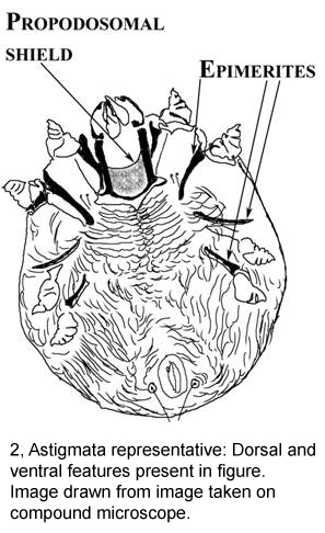
| 11. | At least legs I and II epimerites (Fig 23.) are present and sclerotized. Palp (Fig 24.) dorsally with at least 4 obvious segments (habitus: Fig 61.). | Cheyletiellidae (Prostigmata) |
| - | Epimerites not present. Palp (Fig 25.) dorsally with two or three obvious segments at most (habitus: Fig 63. in part) (body length 200 – 685µm). | Harpirhynchidae (Prostigmata), in part. |
| 12. | Sternal shield (Fig 26.) very noticeable and strongly sclerotized, wider than long. Genital plate (Fig 27.) arrowhead shaped much longer than wide and terminates in blunted point, peritremes extend (Fig 26.) to at least mid-coxa II. Single continuous dorsal shield present (habitus: Fig 57.) (body length 750 – 1040µm) Single species recorded from North American birds Ornithonyssus sylviarum (Canestrini and Fanzago). | Macronyssidae (Mesostigmata) |
| - | If sternal shield (Fig 28.) very noticeable and strongly sclerotized then either: (a) genital shield (Fig 29.) not arrowhead shaped, instead more tongue shaped with rounded terminus, never pointed; or (b) if genital shield is arrowhead shaped then peritremes (Fig 30.) absent or do not extend to mid-coxa II or if they do extend to mid-coxa II then two dorsal shields present (see also Mesostigmata on Representative Page). | 13 |
| 13. | Sternal shield (Fig 31.) very noticeable and strongly sclerotized with at least 3 pairs of sternal setae on sternal shield. Sternal shield approximately as long as wide. Cheliceral digits (Fig 32.) with obvious dentition (habitus: Fig 56.) (body length 970 – 1050µm) . | Laelapidae (Mesostigmata) |
| Laelapidae: Single genus recorded from Alberta, Androlaelaps, distinguished by basally inflated pilus dentilis (Fig 33.) on the fixed cheliceral digit. | ||
| - | Sternal shield either easily discernable or not. If sternal shield (Fig 34.) easily discernable then may be at least twice as wide as long with maximum of two pairs of setae on shield. Cheliceral digits (Fig 35.) without obvious dentition. | 14 |
| 14. | Tritosternum (Fig 37.) present. Peritreme present (Fig 36.), usually extends beyond the anterior margin of coxa III. If peritreme does not extend beyond anterior margin of coxa III and tritosternum present then propodosomal and hysterosomal shields are joined yielding a single shield. Well sclerotized mites. Sternal shield (Fig 34.) strongly sclerotized, wider than long, with 1 or 2 pairs of setae and 0-2 pairs of pores. Chelicerae (Fig 38.) highly elongate distal segment, and minute moveable digit (habitus: Fig 55.); (body length 710 – 1100µm) | Dermanyssidae Dermanyssus (Mesostigmata) |
| - | Tritosternum usually absent. If tritosternum present then dorsal and ventral body setae distally inflated, or sternal shield is either lightly sclerotized and longer than wide, or shield absent. If peritreme (Fig 39.) present it usually does not extend beyond the anterior margin of coxa III, if peritreme does extend beyond the anterior margin of coxa III then tritosternum absent. Peritreme (Fig 40.) may be absent. Poorly sclerotized mites. (habitus: Fig 58.) | Rhinonyssidae (Mesostigmata) |
Key to the Genera of Blood and Tissue Feeding Mites of Ereynetidae, Cheyletiellidae, and Rhinonyssidae, Known from Albertan Birds.
Ereynetidae (Prostigmata):
| 1. | All idiosomal and leg setae barbed (Fig 41.) (body length 500 – 515µm). | Boydaia |
| - | Idiosomal and leg setae (Fig 42.) not all barbed (body length 490 – 550µm). | Neoboydaia |
Cheyletiellidae (Prostigmata) (females only):
| 1. | Hysterosomal shield (Fig 43.) present, may be faintly sclerotized (body length 340 – 390µm). |
Ornithocheyletia
|
| - | Hysterosomal shield absent. | 2 |
| 2. | Palpal claws (Fig 44) and tarsi I-IV claws very small, propodosomal shield (Fig 45.) distinct and without striations (body length 475µm). | Bakericheyla |
| - | Palpal claws (Fig 46.) and tarsi I-IV claws relatively large, propodosomal shield (Fig 47.) indistinct and lined with striations(300 – 570µm). | Neocheyletiella |
Rhinonyssidae (Mesostigmata) (females only):
| 1. | Stigma with peritreme (Fig 48.). | 2 |
| - | Stigma without peritreme (Fig 49.). | 3 |
| 2. | Chelicerae with both digits (Fig 50.), cheliceral shaft distally attenuated (Fig 51.) (body length 670 – 1410µm). | Ptilonyssus |
| - | Chelicerae without fixed digit (Fig 52.), cheliceral shaft with uniform diameter (body length 1100 – 1400µm). | Rhinoecius |
| 3. | Cheliceral shaft distally attenuated (Fig 53.), digits minute (body length 510 – 900µm). | Sternostoma |
| - | Cheliceral shaft approximately uniform diameter (Fig 54.), digits robust (body length 1100 – 1330µm) | Rhinonyssus |
Figure Captions:
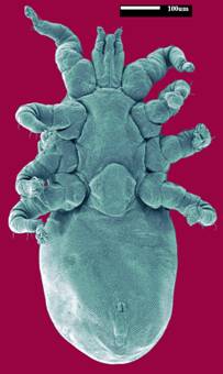
1, Ixodida representative: Dorsal and ventral features present in figure. Image drawn from image taken on compound microscope. 2, Astigmata representative: Dorsal and ventral features present in figure. Image drawn from image taken on compound microscope. 3 & 4, Prostigmata representative: Image redrawn from Fig. 18 Ornithocheyletia gersoni Smiley, from Smiley (1970). 5, 6 & 7, Mesostigmata representative: Image redrawn from Fig. 7, Dermanyssus hirundinis (Hermann), from Moss (1968). 8, Ixodida hypostome. 9, Ixodida Haller’s organ on tarsus 1. 10, Ixodidae prodorsal shield. 11, Mesostigmatan stigmata. 12, Prostigmatan peritreme. Image by G. Williams and W. Knee. 13, Trombiculid trichobothria and prodorsal shield. 14, Syringophilid whole mount. 15, Syringophilid empodium on tarsus 1. 16, Ereynetid barbed leg setae. 17, Ereynetid ereynetal organ in tibia 1. 18, Ereynetid reticulate cuticle on leg. 19, Harpirhynchid whole mount with asymmetrically short legs 3 and 4. 20, Harpirhynchid gnathosoma showing serrate setae distally on palp. 21, Turbinoptid tarsal claws legs 1-4. 22, Knemidokoptid propodosomal shield and pattern of cuticle on dorsum. 23, Cheyletiellid whole mount showing epimerites. 24, Cheyletiellid gnathosoma showing palpal segmentation. 25, Harpirhynchid gnathosoma showing palpal segmentation (below). Harpirhynchid whole mount (above). 26, Macronyssid showing sternal shield, peritreme and stigma. 27, Macronyssid female genital shield. 28, Laelapid female showing genital and sternal shields. 29, Rhinonyssid female genital shield. 30, Rhinonyssid peritreme. 31, Laelapid sternal shield. 32, Laelapid chelicerae. 33 Androlaelaps (Laelapidae) female chelicerae. 34, Dermanyssus (Dermanyssidae) sternal shield. 35, Ptilonyssus (Rhinonyssidae) cheliceral digits. 36, Dermanyssus (Dermanyssidae) peritreme and stigma. 37, Mesostigmatan gnathosoma showing the tritosternum. 38, Dermanyssus (Dermanyssidae) chelicerae. 39, Rhinonyssid peritreme. 40, Rhinonyssid stigma without a peritreme. 41, Boydaia (Ereynetidae) tarsus showing barbed setae. 42, Neoboydaia (Ereynetidae) leg 2 (above) and leg 3 (below) showing setiform setae. 43, Ornithocheyletia (Cheyletiellidae) dorsal opisthosoma showing hysterosomal shield. 44, Bakericheyla (Cheyletiellidae) showing tarsal claws on the palp, and legs 1 and 2. 45, Bakericheyla (Cheyletiellidae) propodosomal shield. 46, Neocheyletiella (Cheyletielldae) showing tarsal claws on the palps and legs. 47, Neocheyletiella (Cheyletiellidae) propodosomal shield. 48, Rhinonyssid stigma with a peritreme. 49, Rhinonyssid stigma without a peritreme. 50, Ptilonyssus (Rhinonyssidae) cheliceral digits. 51, Ptilonyssus (Rhinonyssidae) cheliceral shaft. 52, Rhinoecius (Rhinonyssidae) chelicerae. 53, Sternostoma (Rhinonyssidae) chelicerae. 54, Rhinonyssus (Rhinonyssidae) chelicerae. 55, Dermanyssid whole mount. 56, Laelapid whole mount. 57, Macronyssid whole mount. 58, Rhinonyssid whole mount. 59, Knemidokoptid whole mount. 60, Turbinoptid whole mount. 61, Cheyletiellid whole mount. 62, Ereynetid whole mount. 63, Harpirhynchid whole mount. 64. Trombiculidae whole mount. 65, Feather mite (Astigmata) tarsus and ambulacrum. 66, Nasal mite (Turbinoptidae: Astigmata) legs 1 and 2. 67, Metanalges (Analgidae) male, Psilobrephosceles (Alloptidae) male, and Grallobia (Pterolichidae) female, feather mites. Images by H. Shannon, and W. Knee. 68, Mesostigmatan male, showing gonopore located anteriorly on the sternal shield. 69, Mesostigmatan male showing the spermadactyl fused to the moveable digit. 70, Mesostigmatan female genital shield. 71, Mesostigmatan female chelicerae.
![]()
Biological Survey of Canada
Commission biologique du Canada