
Diagnostic notes
-This species has 6 spines per side on the elytral declivity, and is very similar to I. calligraphus.
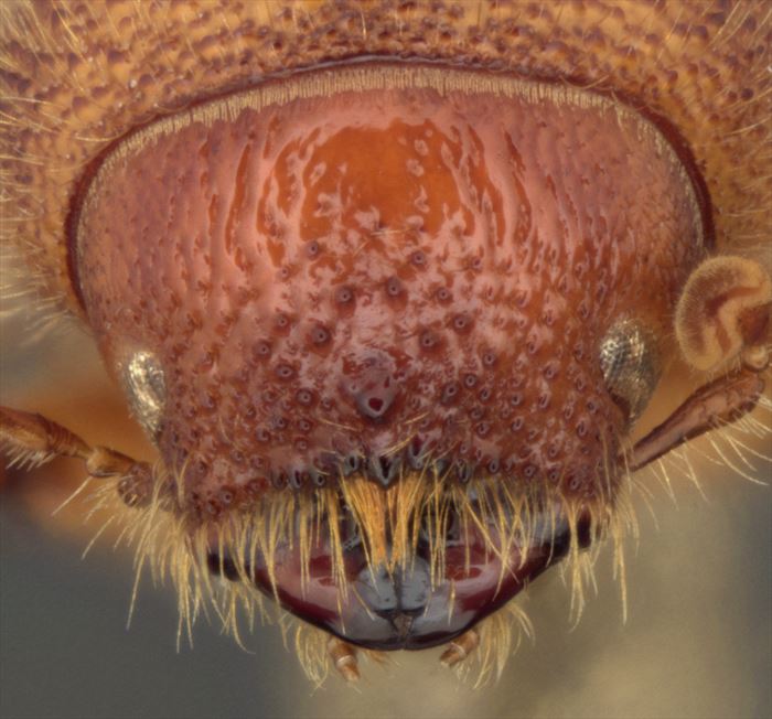
–Frons central tubercle is above the epistomal margin by a distance of twice its width or more in male I. apache but less than twice its width in male I. calligraphus ponderosae.
-Elytral interstrial punctures wider than 0.5 the width of strial punctures in I. apache but less than 0.5 the width of strial punctures in I. calligraphus calligraphus.
–I. apache is often smaller (pronotal width 1.3 to 2.1 mm) than I. calligraphus (pronotal width 1.3 to 2.5 mm) and striations on the pars stridens are narrower (Mean values: I. apache = 0.9 µm; I. calligraphus = 1.0 µm) (Lanier et al. 1991).
Morphological Summary
females
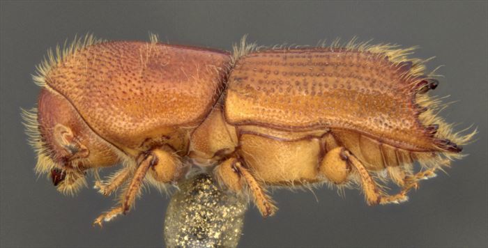
Body. 3.8-5.3 mm long, 2.6-2.9 times longer than wide; pronotum 1.2-1.4 times longer than wide.
Head. Epistomal margin with uniseriate row of tubercles with elongate mesal tubercle. Frons outline convex; vestiture fine – not hiding part of integument; surface sculpture near epistoma densely tuberculate-punctate; central carina present or absent; central tubercle present and single, separated from base of epistomal setae by 0.5-4(-5) tubercle diameters, without pair of circular tubercles on either side of midline; transverse carina absent or present; circular tubercles above top of eyes present – up to, or more than one third of all granules. Vertex and pronotum with stridulatory apparatus (pars stridens). Antennal club sutures acutely angulate.
Prothorax. Protibiae with four socketed teeth on apical half (does not include apical spine).
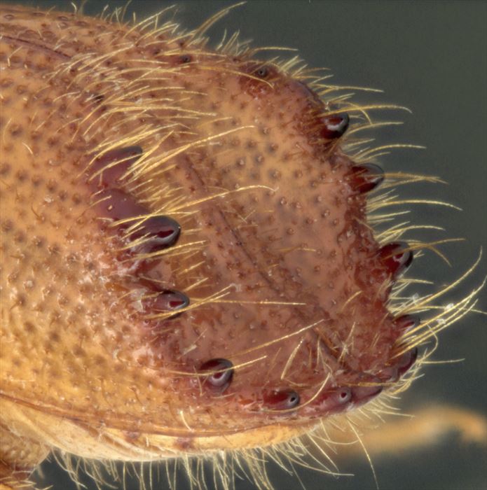
Elytra. Interstriae impunctate (observed on interstriae 2 and 3 on middle third of elytral disc), punctures 0.5-0.8 times diameter of adjacent strial punctures (punctures and striae measured at steepest part of puncture wall), interstriae 2-3 times as wide as adjacent striae. Elytral declivity with six spines per side, spine 3 largest; spine 1 (largest on 2nd interstria) closer to suture than spine 2; spines 1 and 2 separated at base by distance greater than height of spine 1; spine 2 closer to spine 3 than spine 1; spine 3 straight sided with tapered apex, apex acute or right-angled, with apical half symmetrical in lateral view; spines 2 and 3 on or not on shared tumescence, not in line with spines 1 and 4 (posterodorsal view); spine 4 closer to spine 3 than spine 5; declivital integument shiny.